


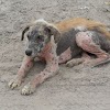
Broken
horn injury


Introduction:
Horns
are the weapon of animals. Anatomically, horn is extension of corneal process
of frontal bone. Broken horn injury is the condition manifested by the any
injury on horn that causes injury to the horns of animals. Mainly long horns of
bulls and goats have the highest possibility of broken horn injury due to
fighting in enclosed place or striking their horn against solid wall. Mostly on
farm this condition is not even noticed sometimes. This condition was mostly asymptomatic expect for
visual presence in few cases. Broken horn
repairs must be done as soon as possible after the break. Most horns
break and go down rather than up, therefore the repair must normally lift the
horn back up to a normal symmetrical shape. Often the horn will not go back
into the exact original place. It will not easily squeeze into the jagged
edges and will need pressure.
Etiology:
There
are many etiological factors that can lead to broken horn injury,
·
Direct violence during fighting with each others
·
Blow or Striking the horn against wall or fixed object
·
Repeated injury setting up a chronic inflammation
·
Automobile Accident
Clinical signs:
This condition is
mostly asymptomatic expect for visual presence in few cases;
·
Asymmetrical horn as one of them may be broken
·
Bleeding from the horns due to rupture of corneal artery
·
When animal walk broken horn moves and animals tends to walk
slowly
·
Animals do not allow anyone to touch or dress the part
·
Inappetence
·
Slowly loss of weight
·
While touching on horns, animals felt pain
·
Crakes and fissures can be seen on horns
·
Inflammation of affected part
·
Lower direction of one horn while other horn is upright
·
The direction of fracture is usually oblique and broken
surface is more or less irregular
·
Fracture of horn cause hemorrhage into frontal sinus and
bleeding from nose
·
Further complication may lead to purulent sinus and empyema
of sinus
Diagnosis:
Diagnosis
of the broken horns can be made by following ways,
- v History: previous history of
accidents or any records of traumatic injury of animals should be noted in case
that may be the cause of broken horn. Most commonly seen on animals with long
and uncurled horns.
- v Inspection of animal horns:
on normal condition horns of animals are symmetrical and upward directed
(commonly). On close inspection we can see crakes and fissures on horns.
- Radiography: x-ray imaging is important diagnosis for broken horn.
- Differential diagnois:
- Horn cancer: It is a
squamous cell carcinoma of the horn, most commonly affecting bullocks. The
disease is restricted to old animals mostly long horned, white coat breed of
cattle.
- Avulsion of horn: It refers to separation of horny covering from the bony core.
- Sinusitis: it is often common sequel to dehorning in animals. During amputation bone sawdust may enter sinus that cause irritation and infection with purulent discharge.
- Firstly cleaning of the affected, exposed horns core and protect it with an antiseptic pad and bandage
- Use of fly repellents and disinfectants to prevent further contamination
- Applying cooling, astringent, antiseptic lotion with a view to arresting hemorrhage
- Apply pressure to the stump
of horn to control bleeding, once bleeding is under control apply a blood stop
powder to encourage clotting and then wrap the horn with bandage
- A temporary tourniquet around the base of horn will stop heavy bleeding but not for long period of time. Release it on every 20 minutes time interval.
- Dehorning: it can be done as a
prophylactic treatment before injury happens or after injury depending of part
of horn that gets broken.
x
Treatment:
Procedure-
§ Hair around the base of buds
are clipped and site should be prepared aseptically
§ Vaseline should be applied
all around the base of horn bud and eye should be covered with clean cloth
§ Potassium or sodium
hydroxide stick is applied with firm pressure in circular fashion till oozing
of blood occurs
§ Or horn bud is removed with
help of sharp knife along with 0.25cm wide piece of skin with base of button.
The haemorrhage checked by digital pressure.
Casting: It used when there is incomplete fracture, crakes or fissures on horn so that they can attach
later.
§ If it is broken and
dirty it becomes less likely that the repair will be successful. Go ahead
and wash out the dirt and proceed.
§ Bend the rebar to the
shape of the top side of the horn spread.
§ Roll the soft cloth in a ball and place
them right on top of the pole. Place the rebar on top of the cloth.
Start wrapping the casting material around the good horn which will pull the rebar up above the bad horn.
roll (rolls are about 4 foot long) around the rebar and good horn all the way from the head to the end of the rebar.
The casting material will dry in a few minutes. Start a second roll of casting material around the broken horn and draw it up to the rebar (like a spring) to pull it up into correct placement.
Wrap it from the head to the end of the rebar. Then take 2 or 3 more rolls and figure eight it back and forth all over the top of the head connecting all the material together. Totally cover all the rebar and cloth tight.
Amputation of horn: is indicated in adult animals following irreparable injuries of horn. Most commonly used technique is flap method of amputation of horn described by Angelo and Das (1970), as it is simple and superior because of less haemorrhage, pain and short healing period.
1. The procedurer cornual nerve block with 2% lignocaine hydrochloride to reduce pain and ligation of cornual artery for bloodless operation.
2. The hair around the base of horn should be clipped and cleaned.
3. An elliptical incision is given around the base of horn and skin on both sides of incision is reflected to form a flap. The incision should be 0.5 cm from horn skin junction.
4. Then vessels are located in temporal fossa by blunt dissection. Haemorrhage is controlled by point firing or using potassium permanganate crystals.
5. The exposed horn is now cut at its bottom with a wire saw. The wire saw produce appreciable heat as a result of friction which results further haemostasis.
6.The skin flaps are apposed
together by interrupted or mattress suture.
7. A protective bandage is
applied after covering the wound.
8. On the ligature is removed on the next day and aseptic dressing is employed.
9. The post treatment period is comparatively shorter.

chandrakala rana magar
(8th semester)
(institute of agriculture and animal science)


0 Comments