


Manoj karki (BVSC&AH)
Date: 2020/5/19
Date: 2020/5/19

Canine demodicosis is a
severe parasitic skin disease of a dog caused by overpopulation of the
follicular mite of various demodex species. One of the most common cutaneous
infection encountered in canine practice is demodicosis.
Demodex mites are normal flora localized in the skin of most apparently healthy dog, when Secondary bacterial infection of the hair follicle often occurs, and rupture of the hair follicle wall may lead to presence of free mites in the dermis and a severe pyogenic infection. Although, demodex canis is acquired by puppies within the first few hours of suckling it is a normal inhabitant of canine skin and demodicosis is not a contagious disease.

Fig: 1.
Life cycle of Demodex canis: (1) the life cycle of Demodex includes,
apart from the adult mite, egg, larva, and two nymphal stages; (2) the
lifecycle happens in hair follicles. The puppy is infected by the dam during
the first days of its life. The infestation is preceded by the multiplication
of the mites on the dam’s skin (3). The mechanism, which accelerates the
multiplication just before whelping, is not known. Most dogs harbor single,
latent Demodex mites in their hairfollicles, living in quiet
seclusion. In some individuals and in some circumstances the mites can start to
multiply uncontrollably, leading into the symptomatic demodicosis.
Etiology:
Demodex canis
is the main causative agent of canine demodicosis. The mites residue in the
hair follicle and sometimes the sebaceous gland. Four stage are seen, the
diamond shaped egg, the 6 legged larvae and 8 legged nymph form which develops
into adult.
Generally three types
of Demodex mites are found in dog. Demodex canis (long bodied mites), Demodex
injai (large bodied mites) and Demodex cornei (short bodied mites)
 |
| Fig: 2. Demodex injai (10×magnification) |
 |
| Fig: 3. Demodex canis and Demodex cornei (blue box) at 10× magnification. |
Pathogenesis:
The mites enter the
hair follicle, reach the root and multiply. Due to inflammation the papilla is
destroyed and the hair is lost. The parasite then enter the sebaceous gland
where condition appear to be very congenial for the growth of mites, since the altered
sebum is a very favourable medium for them to grow. The sebaceous gland may be
dilated and become cystic and lined by squamous epithelium. Thus the skin comes
to be covered by scaly material which may be desquamated. The sebaceous cyst
may rupture, the spilled sebum causing inflammation locally. The blood vessels
are congested and dilated.
Clinical
sign:
The most common
clinical sign are alopecia, scaly-crustly thickened skin with hyperpigmentation, leechinification, erythema,
pruritus and seropurulent discharge seems mostly on per ocular area, face, neck,
shoulder and fore quarter region. Anemia may also occur in this condition due
to loss of skin proteins and leukocytosis. Most case of demodecosis are
non-pruritus unless there is secondary pyoderma. Very rare ulceration may
develop, especially on the face and mucocutaneous area which may mimic
autoimmune disease.
Canine demodectic mange
is classified as two forms.
Localized and Generalized form
Localized
form:
The localized form typically starts as one or more focal alopecic lesion,
erythema and comedomes in dog less than 1.5 years old. Usually only the head
(perioral, peri ocular) or the forelimb are involved. There is no significant pruritus. This is
commonly seen in pups of 3 to 9 month old and spontaneous recovery without
treatment.
Generalized
form: Generalize
demodicosis is a severe disease requiring aggressive therapy. This form
involves the large part of the dogs skin and bears a guarded prognosis.
Generalized form is however, characterized by number of area of localized
disease or even infection in entire skin areas. Generalized demodicosis can be
severe often complicated with secondary bacterial infection and life
threatening.
It should be remember
that every case of generalized demodicosis was localized form.
Juvenile onset
demodicosis (onset prior to puberty) and adult onset demodicosis
Juvenile onset is by far the most common and although a serious disease, often a better prognosis than does adult onset. Adult onset is usually associated with severe internal disease and Is often very difficult to control.
 |
Fig:3. Dog with lesions at face and neck regions
|
 |
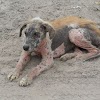
Fig: 4. Alopecia and scale/crusts
formation throughout the body due to infestion of demodectic mite
|
Diagnosis:
Canine demodicosis is
usually diagnosis by identifying mites in skin scrapping and hair plugs.
Performing of skin
scrapping:
1. Some
3-5 site are selected for skin scrapping
2. The
hair, if present, is clipped
3. The
skin is gently squeezed between thumb and forefinger to force the mites more
superficially in the hair follicle.
4.The
skin is moistened with liquid paraffin or mineral oil
5. Some
is also placed on the slide
6. The
skin is then scraped using a blunted scalpel blade until capillary bleeding is
observed.
7. The
material is transferred to the slide
8. The
entire slide is scanned using 10X objective
9. Focus
on suspicious area using the 40Xobjective if necessary.
10. The
proportion of live and dead mites of adult and young forms and of eggs should
be recorded.
or
The
scraping was placed in the test tube and 10% KOH solution was added. Then, the
solution was gently heated (near up to the boiling) with frequent shaking for
about 5-10 min until all the debris was digested. Often the solution was
allowed to cool for some time and was allowed to centrifuge at 2000 rpm for
10min, supernatant were discarded and remaining sediment were a coverslip. The
sediment was observed for mites with the help of compound (10x) microscope.
Prognosis:
The prognosis for juvenile onset localized
demodicosis is very good. The prognosis for juvenile for onset generalized
demodicosis is always guarded.
The
demodicosis mange upto 1 year of age were more susceptible to infection and the
highest prevalence of the disease was found in winter season rather than summer
or rainy season. Demodicosis is highly prevalent in dog whose immune system is
distributed or not well developed. So it is highly recommended that dog should
be given proper nourishment, immunization, deworming and care to avoid skin
disorder like demodicosis.
Treatment and
prevention:
Treatment should be
occur in localized and generalized demodicosis
In localized, this is
usually a self-limiting disease that cures spontaneously. Treatment with
parasiticides is usually not warranted. But 10 % of case of localized
demodicosis go on to become generalized. Therefore whether or not treatment is
given a careful follow up is necessary.
In generalized
demodicosis, it can be one of the most frustrating skin disease, one will ever
treat.
Specific treatment of
demodectic mange are:
Amitraz: 0.025%
whole body dip applied every 2 weeks interval 5-8 application or till skin
scraping are negative. Not recommended for cat and puppy
Ivermectin: 0.3-0.6
mg/kg bwt PO single dose
Moxidectin: 0.4mg/kg
bw POsingle dose
Before application of
medicine crust, scale and debris should be removed with soap and shampoo.
A course of antibiotic
should be given to inhibit bacterial infection. Antibiotic therapy should be
continued until the mite population is well controlled.
Reference:
1. Ashfaq, K., et al. (2019). “Alternative therapeutic approach to treat
canine demodicosis”. EC veterinary science 4.4 :251-256
2. Islam, M.M., et al. (2000). “Prevalence
and pathology of demodectic mange in stray dog in Bangladesh”. Journal of
science and technology. 11: 118-121
3. Shrestha, D., et al. (2015). “Prevalence
of demodectic mange in canine of KTM valley having skin disorder and its
associated risk factor”. Int. Appl sci biotechnol, vol 3(3): 459-463
4. Salem, N.Y., et al. (2020). “Canine
demodicosis hematological and biochemical alteration”. Veterinary
world.org., vol(13):68-72
5. Bhatia, B.B., et al.(2016). “ text book
of veterinary parasitology”. 4th edition.
6. Picture are taken from google.


0 Comments